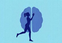
Pain is a universal experience, but the way it arrives in consciousness is anything but simple. At first glance, pain seems like a straightforward alarm: something hurts, your brain tells you to react, and you pull your hand away. But underneath that simple reflex is a vast orchestra of cells, chemicals, pathways, and interpretations. The brain doesn’t just receive pain signals; it decodes them, filters them, ranks them by importance, colors them with memory and emotion, and sometimes, frustratingly, generates pain with little or no warning. In this article we’ll walk step by step through how the brain processes pain signals, explore the key players from receptors to the cortex, and discuss what happens when the system goes awry. Expect clear explanations, real-world examples, lists that simplify complex ideas, and a handy table that ties brain regions to their roles in pain processing. Whether you’re curious about neuroscience, living with pain, or caring for someone who is, this guided journey will make the mechanisms behind pain make sense.
Содержание
First Contact: How Pain Begins at the Periphery
Pain typically begins outside the brain, at the very tips of our body where specialized nerve endings sense danger. These endings are called nociceptors — “noci” meaning harm. Nociceptors are tiny sensory neurons distributed through skin, muscles, joints, and internal organs. They don’t fire continuously; instead they remain quiet until confronted with potentially damaging stimuli such as intense heat, mechanical pressure, or chemical irritants. When triggered, nociceptors translate physical or chemical forces into electrical impulses: the language the nervous system understands.
There are several types of nociceptors. Some respond to heat, some to mechanical pressure, and others to inflammatory chemicals released during tissue damage. A single injury can activate multiple nociceptor types simultaneously, producing a mixed signal. That mixed signal is important because it helps the brain figure out what kind of injury occurred — a sharp cut, a burn, a sprain — and how urgently it needs to act.
These primary sensory neurons have long fibers called axons that carry the electrical signal into the spinal cord. The speed and pattern of these impulses matter: a fast, sharp pain is usually carried by myelinated A-delta fibers, while longer-lasting, dull aching pain travels on slower, unmyelinated C fibers. This difference in fiber type explains why a stubbed toe often produces a quick sharp pain followed by a lingering throb.
Types of Peripheral Pain Signals
The diversity of peripheral signals helps the brain tailor its response. Here are the major categories:
- Mechanical nociception: caused by cutting, crushing, or stretching tissue.
- Thermal nociception: caused by extreme heat or cold.
- Chemical nociception: caused by irritants, or by inflammatory molecules released after injury.
- Polymodal nociception: nociceptors that respond to a combination of the above.
This layer of complexity at the periphery sets the stage for all downstream processing. The brain receives not just an “ouch” signal but rich contextual data about what might be wrong.
Entry Gate: The Spinal Cord and Dorsal Horn Processing
Once nociceptors generate electrical impulses, those signals enter the spinal cord through dorsal root ganglia and terminate in the dorsal horn — the spinal cord’s traffic control center for sensory inputs. The dorsal horn is a dense network of neurons that sort and modulate incoming messages before forwarding them upward. It’s here that the first real processing and filtering of pain signals occurs.
Neurons in the dorsal horn do more than relay signals — they integrate inputs from multiple sources. Incoming pain fibers meet inhibitory interneurons, excitatory projection neurons, and descending inputs from the brain that can either amplify or suppress the message. Think of the dorsal horn as a security checkpoint: it checks the signal’s credentials and decides whether to let it pass, hold it, or dampen it.
This checkpoint function is crucial because everyday life would be impossible if every minor irritation produced a full alarm in the brain. The spinal cord’s ability to gate signals — a principle known historically as the gate control theory of pain — helps prioritize threats. For example, rubbing a bumped elbow often reduces pain because touch fibers engage inhibitory circuits in the dorsal horn that partially block incoming nociceptive signals.
Key Cellular Players in the Dorsal Horn
Several cell types shape spinal pain processing:
- Projection neurons: send pain signals up to the brain via ascending tracts.
- GABAergic and glycinergic interneurons: provide inhibitory control to reduce signal transmission.
- Excitatory interneurons: amplify certain signals during inflammation or injury.
- Microglia and astrocytes: non-neuronal cells that can modulate pain through immune-like responses, especially in chronic pain.
When the balance between excitation and inhibition shifts toward excitation — due to severe injury, inflammation, or persistent stimulation — the dorsal horn becomes hyperexcitable. That hyperexcitability is a major contributor to chronic pain conditions and to phenomena like referred pain and allodynia (when light touch becomes painful).
Ascending Highways: How Signals Climb to the Brain
From the dorsal horn, projection neurons launch the pain signal upward along major ascending pathways. The most famous of these is the spinothalamic tract, a direct route that carries nociceptive information to several brain regions, including the thalamus. The spinothalamic tract is often described as the “fast lane” for pain, but it’s not the only route. Other ascending tracts reach brainstem nuclei and the reticular formation, which are involved in arousal and autonomic responses.
Along these ascending pathways the signal branches, allowing parallel processing. One branch heads to the thalamus and then to the primary and secondary somatosensory cortices, which map the body and encode pain location, intensity, and quality. Another branch goes to deeper brain structures (like the periaqueductal gray, limbic system, and hypothalamus) that contribute to emotional responses, memory, and behavioral reactions. This parallel routing is why pain is simultaneously a sensory and an emotional experience: your brain learns where it hurts, how badly it hurts, and what it means.
Spinothalamic Tract and Beyond
Key destinations for ascending pain signals include:
- Thalamus: the main relay station that directs signals to cortex.
- Somatosensory cortex: encodes the physical attributes of pain (where and how strong).
- Insula and anterior cingulate cortex (ACC): interpret internal bodily states and emotional aspects.
- Brainstem nuclei: regulate automatic reactions like heart rate and breathing.
Because the pathway is distributed, damage or modulation at various points can alter pain perception. For instance, certain strokes affecting the thalamus can produce severe chronic pain syndromes, highlighting how important central processing is for the experience of pain.
The Thalamus: Central Relay and Filter
The thalamus sits deep within the brain and acts as a hub for nearly all sensory information. For pain, it’s not a passive relay but an active processor. Different thalamic nuclei receive pain-related inputs and forward them to specific cortical regions. The thalamus can amplify signals, filter them, or gate them based on context and arousal state. During sleep, for example, thalamic gating reduces the likelihood that minor painful stimuli will wake you. In contrast, when you’re alert and anxious, thalamic activity can heighten sensitivity to the same stimulus.
Besides gating, the thalamus plays a role in synchronizing cortical networks so that the brain can build a coherent representation of pain that includes sensation, emotion, and potential actions. This synchronization is mediated by rhythmic activity in certain frequency bands, which can increase during painful experiences.
Thalamic Modulation in Chronic Pain
In chronic pain conditions, thalamic function can become altered. Some patients display abnormal thalamic rhythms and heightened thalamic responsiveness, which may perpetuate the chronic pain state. Understanding thalamic dysfunction has practical implications: treatments like deep brain stimulation targeted to thalamic nuclei are explored for intractable pain.
Cortical Processing: Where Pain Becomes Experience
When pain signals arrive in the cortex, they’re processed across a distributed set of regions often called the “pain matrix.” This network includes the primary somatosensory cortex (S1), which maps the body surface; the secondary somatosensory cortex (S2), involved in integrating bilateral sensations; the insula, which monitors internal bodily states; the anterior cingulate cortex (ACC), associated with emotional and motivational aspects of pain; and prefrontal areas involved in cognitive evaluation and decision-making.
The somatosensory cortices answer the question “where and what” about pain. They allow you to pinpoint the painful spot and to discriminate qualities like sharp versus burning. The insula and ACC answer “how it feels and what it means” — whether the pain is distressing, whether it feels urgent, and whether it should trigger avoidance. Prefrontal regions that analyze past experiences and future consequences influence how you interpret pain and how you choose to respond. Together, these cortical computations create the subjective experience we call “pain.”
Integration and Conscious Awareness
One of the fascinating aspects of cortical processing is that conscious awareness of pain emerges through integration, not simply by activation of a single brain region. Pain is a constructed experience — the brain combines signals about intensity and location with context (Are you injured? Is this expected?), memories (Did this feel like something that healed before?), and emotions (Are you fearful or calm?). That’s why two people can have very different pain experiences from the same injury.
Neural plasticity in cortical regions can also change long-term sensation. Repeated painful experiences remodel synapses and can enlarge or shrink cortical representations of body parts, which is one mechanism implicated in chronic pain and phantom limb pain.
Neurochemistry of Pain: The Messengers that Make It Hurt
Pain processing depends on a variety of chemical messengers — neurotransmitters and neuromodulators that excite, inhibit, or modulate neuronal activity. At the synapses of nociceptive pathways, glutamate is the principal excitatory neurotransmitter, rapidly transmitting pain signals. Substance P and calcitonin gene-related peptide (CGRP) are neuropeptides that sustain and amplify pain signals, especially during inflammation.
Inhibitory neurotransmitters like GABA and glycine tone down pain transmission in the spinal cord and brain. Monoamines such as serotonin and norepinephrine are part of descending pathways that modulate spinal processing; their levels and receptor activity can substantially affect pain sensitivity. Endogenous opioids — endorphins, enkephalins, and dynorphins — act within the brain and spinal cord to dampen pain perception and are the body’s built-in analgesic system.
How Drugs Target These Chemicals
Understanding pain chemistry explains many treatments. Opioid drugs mimic endogenous opioids to blunt pain, but their side effects and risk for addiction are major clinical concerns. Antidepressants that increase serotonin and norepinephrine can strengthen descending inhibition and are commonly used for some chronic pain conditions. Nonsteroidal anti-inflammatory drugs (NSAIDs) reduce peripheral inflammation and lower chemical activation of nociceptors. Emerging therapies target specific neuropeptides (like CGRP antagonists for migraine) or ion channels that control neuronal excitability.
Descending Control: The Brain’s Pain Brake
The brain doesn’t just passively receive signals — it actively controls and modifies them. Descending pain modulation involves pathways that originate in the cortex and brainstem and project back down to the spinal cord. Key nodes include the prefrontal cortex, periaqueductal gray (PAG) in the midbrain, the rostral ventromedial medulla (RVM), and spinal inhibitory circuits. These systems can reduce pain (analgesia) or, paradoxically, enhance it (facilitation).
The PAG is a central hub: activated by cognitive and emotional factors, it recruits the RVM which then sends descending fibers to the dorsal horn. Depending on the neurotransmitter signals engaged, these pathways release serotonin, norepinephrine, or endogenous opioids to suppress nociceptive transmission. This is why psychological factors like attention, expectation, and stress can alter pain dramatically. For example, intense focus on pain usually amplifies it, while distraction can reduce it through descending inhibitory engagement.
Psychological Influence on Descending Control
Expectations and belief systems strongly sway descending modulation. The placebo effect — where an inert treatment produces real pain relief — operates through endogenous opioid release and modulation of descending pathways. Conversely, negative expectations or fear can activate facilitatory circuits, increasing sensitivity. These effects underline the profound mind-body interactions in pain and why psychological therapies (like cognitive behavioral therapy and mindfulness) can be effective adjuncts for chronic pain management.
When the System Breaks: Chronic Pain and Central Sensitization
Acute pain usually serves a protective purpose and then fades with healing. Chronic pain, however, can persist beyond tissue repair and become a disease in itself. A central mechanism implicated in chronic pain is central sensitization: increased excitability of neurons in the spinal cord and brain that amplifies pain signals and decreases the threshold for activation. Central sensitization can manifest as hyperalgesia (increased pain from a stimulus that normally provokes pain) and allodynia (pain from non-painful stimuli).
Several processes contribute to this maladaptive state: prolonged release of excitatory neurotransmitters, reduced inhibitory tone, neuroinflammation driven by glial cells, and structural synaptic changes that strengthen nociceptive pathways. Over time, pain processing networks can rewire — the brain essentially learns to be in pain. These network-level changes help explain why some chronic pain conditions are resistant to simple symptomatic treatments.
Examples of Central Sensitization in Conditions
Conditions often associated with central sensitization include:
- Fibromyalgia: widespread pain with heightened sensitivity to touch and temperature.
- Chronic low back pain: where central changes maintain pain even after structural issues are treated.
- Complex regional pain syndrome: intense pain and autonomic changes in a limb following injury.
- Phantom limb pain: pain perceived in an amputated limb, reflecting cortical reorganization.
Because central sensitization involves brain and spinal circuits, effective management often requires multimodal strategies addressing both peripheral triggers and central processing.
Individual Differences: Why Pain Is Personal
Pain varies widely between individuals. Genetics, environment, previous experiences, cultural background, emotional state, and attention all shape pain perception. Genetic polymorphisms influence receptors, ion channels, and neurotransmitter systems, altering sensitivity and response to analgesics. Early life experiences and learning shape pain-related memory and threat appraisal. Cultural and social context affects how pain is expressed and managed.
Even gender and age alter pain processing: hormonal differences can influence nociception and modulation, and aging brings changes in pain thresholds and central processing. Clinically, these variations underscore the need for personalized pain management rather than one-size-fits-all approaches.
Psychological factors contribute significantly to pain outcomes. Depression, anxiety, fear-avoidance beliefs, and catastrophizing (expecting the worst) are strong predictors of chronic pain severity and disability. Conversely, resilience, social support, and effective coping strategies can buffer pain and improve recovery. Integrative pain care often includes psychological and social interventions to address these determinants.
Treatments that Target Central Processing
Given the brain’s central role in pain, many effective treatments aim at central mechanisms. These include pharmacological, interventional, rehabilitative, and psychological approaches.
| Treatment | Main Central Mechanism | Typical Use |
|---|---|---|
| Opioids | Activate opioid receptors in brain and spinal cord to reduce transmission | Short-term severe pain; some chronic pain with caution |
| Antidepressants (SNRIs, TCAs) | Enhance descending serotonin/norepinephrine inhibition | Neuropathic pain, chronic widespread pain |
| Anticonvulsants (gabapentin, pregabalin) | Reduce neuronal excitability and synaptic release | Neuropathic pain, some chronic pain syndromes |
| Physical therapy and exercise | Modulate cortical representations and descending control | Musculoskeletal and chronic pain rehabilitation |
| Cognitive behavioral therapy (CBT) | Restructures pain beliefs and engages top-down modulation | Chronic pain management |
| Neuromodulation (TENS, spinal cord stimulation) | Stimulates inhibitory circuits and modifies dorsal horn activity | Refractory neuropathic or chronic pain |
| Mindfulness and meditation | Alters attention, reduces emotional reactivity, engages descending inhibition | Chronic pain adjunct therapy |
No single treatment fits all, and often a combination of approaches — addressing peripheral drivers and central processing — is most effective. Treatment choice is informed by pain type, duration, comorbidities, and patient preference.
Emerging Therapies
Research is exploring new approaches that directly target central mechanisms: drugs that block specific ion channels involved in nociception, monoclonal antibodies against pain-mediating peptides (e.g., CGRP for migraines), and brain stimulation techniques like transcranial magnetic stimulation (TMS) and deep brain stimulation (DBS). Gene therapies and interventions targeting neuroinflammation are also under investigation. These avenues reflect the growing recognition that complex central processes drive many forms of persistent pain.
Putting It All Together: An Everyday Example
Imagine you’re walking barefoot and step on a broken shell. The initial sharp pain comes from A-delta fibers telling the dorsal horn, “This is acute, withdraw now.” You yank your foot back. While you hop on one leg, C fibers keep firing, producing a dull ache that signals tissue damage and prompts you to inspect the wound. Your thalamus directs the signal to the somatosensory cortex where you identify the exact location; the insula tells you it feels hot and raw; the ACC adds the distressing emotional tone — you’re worried this might be a deep tear.
If someone nearby reassures you and helps clean the wound, your prefrontal cortex may reappraise the threat and reduce anxiety. Descending pathways may then suppress pain signals, and endorphins released by laughter or gentle distraction reduce the ache. Conversely, if you recall a prior foot injury that led to infection, anxiety might heighten the pain experience. This simple vignette shows how sensory, emotional, cognitive, and social factors interact in real time to shape pain.
Quick Summary List: The Flow of Pain Processing
- Peripheral nociceptors detect harmful stimuli and generate electrical impulses.
- Signals enter the spinal cord, where dorsal horn neurons integrate and modulate them.
- Ascending tracts carry signals to the thalamus and brainstem, branching to multiple targets.
- The thalamus routes information to cortical regions for localization, intensity coding, and emotional appraisal.
- Descending pathways from brain areas modulate spinal processing, either inhibiting or facilitating signals.
- Neurochemical systems (glutamate, GABA, monoamines, opioids, peptides) mediate transmission and modulation.
- Plastic changes and neuroinflammation can create lasting alterations, leading to chronic pain.
These steps are dynamic and overlap — the brain’s processing of pain is interactive and adaptive, sometimes to our benefit, sometimes to our detriment.
Common Misunderstandings about Pain Processing
It helps to correct a few common myths. First, pain intensity is not always proportional to tissue damage — the brain can amplify or suppress pain independently of injury severity. Second, chronic pain does not always indicate ongoing structural damage; much of it can arise from altered nervous system function. Third, pain is not purely physical or purely psychological; it’s a complex blend of sensation, emotion, cognition, and social context.
Understanding these elements reduces stigma and helps patients and caregivers approach pain with more nuance and compassion.
The Future of Pain Research and Treatment
The future promises more targeted, mechanism-based pain treatments. Advances in imaging, genetics, and computational neuroscience are revealing individual pain signatures and neural circuits involved in specific pain disorders. This knowledge could lead to precision medicine approaches where treatments are matched to the patient’s neurobiological profile. Noninvasive brain stimulation and closed-loop neuromodulation systems that respond to real-time neural activity are being refined. Meanwhile, insights into neuroimmune interactions open possibilities for drugs that reduce neuroinflammation without global immune suppression.
Beyond biology, there’s growing recognition that social determinants of health and access to care shape pain outcomes. Future strategies will likely be multidisciplinary, integrating pharmacology, neuromodulation, physical rehabilitation, and psychological care, tailored to the individual.
Practical Takeaways: Living with and Managing Pain
If you or someone you care for deals with pain, several practical principles can guide management:
- Early, comprehensive assessment matters: identify pain type, triggers, and contributing factors.
- Use multimodal strategies: combine medication, physical therapy, psychological approaches, and lifestyle changes.
- Address sleep, mood, and stress: these strongly influence pain sensitivity and recovery.
- Engage in graded activity: gradual, guided exercise can restore function and remodel pain circuits.
- Seek personalized care: what works for one person may not work for another — be open to iterative adjustments.
Education about pain’s mechanisms can empower patients, reducing fear and encouraging proactive coping. Remember that pain treatment aims not only to reduce sensation but also to restore function and quality of life.
Research Tools: How Scientists Study Pain Processing
Understanding the brain’s pain processing relies on many experimental tools. Human studies use imaging (fMRI, PET), electroencephalography (EEG), and psychophysical testing to map responses. Animal models allow cellular and molecular dissection of pathways and testing of new treatments. Genetic techniques, optogenetics, and chemogenetics permit precise manipulation of specific neurons to observe causal effects. Each tool has limits: findings in animals don’t always generalize to humans, and imaging shows correlates but not causal mechanisms. However, converging evidence across methods steadily advances our understanding.
Short Table: Key Brain Regions and Their Pain Roles
| Region | Primary Role in Pain |
|---|---|
| Primary somatosensory cortex (S1) | Localization and intensity coding |
| Secondary somatosensory cortex (S2) | Integration of bilateral and complex sensory data |
| Insula | Interoception and subjective feeling of pain |
| Anterior cingulate cortex (ACC) | Emotional and motivational components |
| Thalamus | Relay and filter for sensory information |
| Periaqueductal gray (PAG) | Descending pain modulation hub |
| Rostral ventromedial medulla (RVM) | Implicated in both inhibition and facilitation of pain |
This simplified map highlights how widespread and interconnected pain processing is, explaining why it’s rarely solved with single-target therapies.
Ethical and Social Dimensions of Pain
Pain has ethical and social implications. Under-treatment of pain persists in some populations due to disparities in access, biases, or stigma. The opioid crisis has also complicated pain care by highlighting risks associated with certain medications. Balancing effective relief with safety and equity requires systems-level solutions: improved provider training, broader insurance coverage for nonpharmacologic therapies, and patient-centered policies that respect individual needs.
Public understanding of pain biology can decrease blame and increase empathy for people living with chronic pain. Social support and workplace accommodations can be pivotal in helping individuals maintain function and dignity.
Final Thoughts
The journey from a tiny nociceptor firing at the skin to the vivid, multifaceted experience we call pain travels through a landscape rich with neural computation, modulation, and meaning. Pain is not merely a signal; it’s a story the brain tells about the body’s state, past experiences, and future risks. Because pain weaves together sensory, emotional, cognitive, and social threads, its treatment needs to be equally broad and personalized. Advances in neuroscience continue to illuminate the mechanisms behind pain and point to better ways of easing suffering. Through education, compassion, and science-driven care, we can improve outcomes for individuals and communities affected by pain.
Conclusion
Pain arises from a complex dance between peripheral receptors, spinal processing, ascending pathways, thalamic routing, cortical interpretation, and descending modulation, all mediated by a variety of neurotransmitters and shaped by our emotions, memories, and expectations; understanding these mechanisms helps explain why pain can be acute or chronic, why it varies between people, and why effective treatment often requires personalized, multimodal approaches that address both body and brain.

