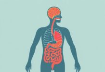
If you’ve ever wondered what makes your thoughts feel immediate, your reflexes lightning-fast, and a pinprick only mildly annoying, you’re already sensing the work of the nervous system. The phrase La Diferencias entre el Sistema Nervioso Central y Periférico captures the heart of a fascinating divide inside your body: two major parts working together but playing very different roles. In this article I’ll walk you through what separates the central nervous system (CNS) from the peripheral nervous system (PNS), why that separation matters, and how both cooperate to make perception, movement, and survival possible.
This is a friendly, conversational tour. I’ll explain structure, cell types, protective mechanisms, how signals travel, what happens when things go wrong, and practical ways to support a healthy nervous system. Expect clear examples, useful analogies, a comparison table, and lists to make the essentials stick. Whether you’re a student, a caregiver, or just curious, you’ll leave with a stronger grasp of how these two systems differ and why both are essential.
Содержание
Why this distinction matters: a short introduction
Think of the nervous system as a communication network. Like any network, it has a central hub and an extensive set of wiring that reaches into every corner. The central nervous system (CNS) — the brain and spinal cord — is like the control center or headquarters. It processes information, forms decisions, and issues instructions. The peripheral nervous system (PNS) is the body’s wiring and the local mail service: nerves that carry messages to and from the CNS and the rest of the body, from fingertips to internal organs.
Understanding La Diferencias entre el Sistema Nervioso Central y Periférico is not just academic. It helps explain why head injuries behave differently from limb neuropathies, why some diseases affect movement while others impact sensation, and why treatments like surgery, drugs, or rehabilitation target different parts of the system. In short, the CNS and PNS are partners with distinct responsibilities; knowing which does what helps diagnose problems and plan care.
Basic anatomy: what each side contains
Anatomy of the Central Nervous System
The CNS has two major components: the brain and the spinal cord. The brain sits protected inside the skull and is the seat of consciousness, memory, emotion, sensation interpretation, and high-level motor control. The spinal cord runs down the spine and is the main highway for signals between the brain and the body. It also handles many reflexes independently of the brain.
At a microscopic level, the CNS is composed of gray matter and white matter. Gray matter contains neuronal cell bodies and is where processing and synapses happen. White matter contains bundles of myelinated axons — the long projections that transmit signals rapidly over distances — creating the communication routes within the CNS.
Anatomy of the Peripheral Nervous System
The PNS contains all nerves and ganglia outside the brain and spinal cord. This includes cranial nerves that leave the brain to serve the head and neck, and spinal nerves that emerge from the spinal cord to serve the limbs and trunk. Peripheral nerves branch extensively to innervate muscles, skin, and internal organs.
The PNS itself splits into the somatic nervous system (controlling voluntary movements and conveying sensory information from skin and musculoskeletal system) and the autonomic nervous system (regulating involuntary functions like heart rate, digestion, and respiratory rate). The autonomic system further divides into sympathetic and parasympathetic branches that often have opposite effects.
How they work: function and signal flow
Signal processing in the CNS
The CNS processes incoming sensory information and transforms it into perceptions, thoughts, memories, and plans. In the brain, different regions specialize — the visual cortex for vision, the auditory cortex for sound, the motor cortex for initiating voluntary movement, and deeper structures like the thalamus or basal ganglia for routing signals and coordinating action. The cerebral cortex, with its folded surface, allows for complex integrative functions such as language and abstract reasoning.
The spinal cord is more than a simple cable. It integrates signals from peripheral nerves and can initiate reflex arcs — quick, protective responses that bypass conscious thought. For example, touching something hot causes immediate withdrawal through spinal circuits before the brain fully interprets the event.
Signal transmission in the PNS
The PNS carries sensory inputs from receptors in the skin, muscles, joints, and organs toward the CNS (afferent signals), and motor outputs from the CNS to muscles and glands (efferent signals). Peripheral nerves are made up of bundles of axons, which may be myelinated (fast conduction) or unmyelinated (slower conduction). Schwann cells, a type of glial cell in the PNS, produce the myelin that insulates these axons.
Because the PNS extends to the body’s periphery, it translates the CNS’s orders into actions — like contracting a muscle or dilating a pupil — and transmits sensations such as pain, temperature, and proprioception back to the CNS. The autonomic branch also uses peripheral ganglia as relay stations to modulate heart rate, blood pressure, and digestion without conscious input.
Cell types and supporting players
The nervous system is not just neurons. Glial cells are crucial supporting actors that maintain the environment, guide development, and assist signal transmission. But the CNS and PNS have different kinds of glia and different arrangements of neurons, which contributes to their distinct behaviors.
Neurons: the signal carriers
Neurons are specialized cells that transmit electrical and chemical signals. They have dendrites to receive inputs, a cell body where signals are integrated, and an axon that carries output to other neurons or effector cells. In both CNS and PNS, neurons use action potentials (electrical impulses) and chemical neurotransmitters to communicate across synapses.
Although neuron function is fundamentally similar, CNS neurons are organized into complex networks densely interconnected in the brain and spinal cord, while many peripheral neurons form more linear pathways linking sense organs or muscles to central processors.
Glial cells: different cast members
In the CNS, the main glial types are astrocytes, oligodendrocytes, microglia, and ependymal cells. Astrocytes help maintain the chemical environment, support the blood-brain barrier, and modulate synaptic function. Oligodendrocytes myelinate multiple axon segments, enabling fast conduction. Microglia act as immune surveillance cells. Ependymal cells line fluid-filled ventricles and help circulate cerebrospinal fluid.
In the PNS, Schwann cells serve a central role by myelinating single axon segments and supporting axon regeneration after injury — a key difference that affects recovery potential. Satellite cells in peripheral ganglia support neuronal cell bodies similarly to astrocytes.
Protection and repair: how each system defends itself
Barriers and armor in the CNS
The CNS is highly protected because damage there can cause profound, often irreversible deficits. Protection comes from three main lines: the skull and vertebral column physically shield the brain and spinal cord; meninges (three layers of membranes) envelope the CNS; and the blood-brain barrier (BBB) selectively filters substances entering the brain from the bloodstream. Cerebrospinal fluid (CSF) cushions the brain and removes metabolic waste.
All these protections, however, come with a tradeoff. The blood-brain barrier restricts immune cell access and some medications, making infections and treatments more challenging. Additionally, neurons in the adult CNS have limited capacity to regenerate after injury, which is why spinal cord and brain injuries are so devastating.
Defense and regeneration in the PNS
The PNS has less rigid barriers, which allows better immune surveillance and easier access for treatments. Peripheral nerves are, however, more exposed to physical injury. The silver lining is that peripheral neurons and Schwann cells create an environment more favorable for axon regeneration. After injury, Schwann cells clear debris and form guiding structures called bands of Büngner, which assist regenerating axons in re-growing toward their targets.
Because of this regenerative capacity, many peripheral nerve injuries—when properly managed—can heal with partial or full recovery, whereas similar damage in the brain or spinal cord rarely regenerates in the same way.
Blood supply and metabolic considerations
Both systems demand energy, but their supply mechanisms and vulnerabilities differ. The brain is metabolically hungry: it uses a significant portion of the body’s oxygen and glucose despite representing only a small percentage of body weight. That makes the brain highly vulnerable to strokes — interruptions in blood supply that cause rapid cell death.
Unique vascular features of the CNS
The CNS blood vessels are tightly regulated by the blood-brain barrier. Endothelial cells are sealed by tight junctions preventing many molecules from entering the brain. While this is protective, it complicates the delivery of many therapies and requires specialized transport mechanisms for nutrients. A stroke or blood vessel rupture can have catastrophic effects because CNS neurons need continuous, tightly regulated blood flow.
PNS blood supply and vulnerabilities
Peripheral nerves receive blood from a network of small vessels in the surrounding tissues. Because there’s less of a barrier, immune cells and systemic factors have more influence on peripheral nerves. This is why systemic conditions like diabetes or autoimmune diseases readily affect peripheral nerves, often causing neuropathy (numbness, tingling, or pain).
Diseases and disorders: how problems differ between CNS and PNS
Some conditions primarily affect the CNS, others the PNS, and some involve both. The location of disease determines symptoms, treatment approaches, and prognosis.
Common CNS disorders
Diseases that primarily impact the CNS include stroke, traumatic brain injury, multiple sclerosis (an immune attack on CNS myelin), Alzheimer’s disease and other dementias, Parkinson’s disease, brain tumors, and spinal cord injuries. Many of these conditions have profound effects on cognition, movement, and autonomic control, and recovery can be limited due to restricted regeneration in the CNS.
For example, multiple sclerosis involves immune-mediated loss of oligodendrocyte myelin in the CNS, leading to patchy deficits in sensation and movement. Treatments often focus on modulating the immune system and rehabilitating lost function.
Common PNS disorders
Peripheral nervous system disorders include peripheral neuropathies (often from diabetes or toxins), Guillain-Barré syndrome (an acute autoimmune attack on peripheral myelin), carpal tunnel syndrome (compression of a peripheral nerve), and peripheral nerve injuries from trauma. Because Schwann cells help support regeneration, some peripheral injuries can recover significantly with time and proper care.
Guillain-Barré is an example of a rapidly progressive peripheral disorder that can cause weakness and paralysis; treatment involves immunotherapy and supportive care. Diabetic peripheral neuropathy develops gradually and causes numbness and pain in a “stocking and glove” distribution, often requiring symptom control and metabolic management.
Diagnostic approaches: how clinicians tell them apart
Distinguishing CNS from PNS problems often starts with the pattern of symptoms. Central lesions may cause weakness or sensory deficits following specific patterns, reflex changes, and cognitive or cranial nerve involvement. Peripheral issues often present with localized numbness, pain, or weakness along specific nerve distributions.
- Imaging: MRI and CT scans are powerful tools for visualizing the brain and spinal cord to find strokes, tumors, or demyelination that affect the CNS.
- Electrophysiology: Nerve conduction studies and electromyography (EMG) evaluate peripheral nerve and muscle function, useful for diagnosing neuropathies and entrapments.
- Lumbar puncture: Sampling cerebrospinal fluid can reveal infections, inflammation, or certain biomarkers pointing to CNS disease.
- Blood tests: These assess metabolic causes like diabetes, autoimmune markers, and infections that may affect peripheral nerves or central structures.
Treatments and recovery: differences in options and prognosis
Therapeutic strategies differ based on whether the CNS or PNS is involved. Because the CNS has limited regenerative capacity and strict barriers, treatments often aim to protect remaining tissue, modulate immune responses, compensate for lost function, or use assistive technology. Rehabilitation (physical, occupational, speech therapy) is crucial to maximize recovery.
Treating CNS disorders
Treatments for CNS conditions include acute measures (clot-busting drugs or thrombectomy for ischemic stroke, surgery for traumatic injuries or tumors), disease-modifying therapies (for example, immunotherapies in multiple sclerosis), and long-term rehabilitation. Experimental approaches, like stem cell therapy and neuroprosthetics, are active areas of research but are not yet routine for large-scale CNS repair.
Because medication must cross the blood-brain barrier, designing effective CNS-targeted drugs is a particular challenge that shapes research priorities and clinical trials.
Treating PNS disorders
PNS treatments often include surgical repair of injured nerves, decompression for entrapment syndromes, immunotherapy for autoimmune neuropathies, and symptomatic treatments (pain control, physical therapy). The greater regenerative potential of peripheral nerves means recovery is often more promising, particularly when timely surgical repair and rehabilitation are available.
For systemic causes like diabetic neuropathy, controlling blood sugar and addressing metabolic risk factors are central to preventing progression and enabling recovery.
Comparison table: central vs peripheral at a glance
Below is a concise table that contrasts key features of the CNS and PNS to make the differences easy to remember.
| Feature | Central Nervous System (CNS) | Peripheral Nervous System (PNS) |
|---|---|---|
| Main components | Brain and spinal cord | Cranial nerves, spinal nerves, ganglia, peripheral plexuses |
| Primary glial cells | Astrocytes, oligodendrocytes, microglia, ependymal cells | Schwann cells, satellite cells |
| Protection | Skull/vertebrae, meninges, blood-brain barrier, CSF | Less rigid protection; nerves embedded in connective tissue |
| Regenerative capacity | Limited; neurons less able to regenerate | Greater; Schwann cells support axon regrowth |
| Typical disorders | Stroke, MS, Alzheimer’s, spinal cord injury | Peripheral neuropathy, Guillain-Barré, entrapment syndromes |
| Diagnostic focus | Neuroimaging, CSF analysis | Nerve conduction studies, EMG, clinical sensory maps |
| Treatment challenges | Drug delivery across BBB, limited regeneration | Managing systemic causes, promoting physical nerve repair |
Everyday examples that illustrate the differences
Concrete examples help solidify the concepts. Here are a few relatable scenarios that show how CNS and PNS problems differ in real life.
- Touching a hot stove: Pain sensors in your finger (PNS) send signals to the spinal cord. A reflex removes your hand before the brain consciously processes the pain (spinal cord, part of CNS). Later you feel the full sensation and understand what happened (brain).
- Stroke vs. neuropathy: A stroke (CNS) might suddenly produce weakness on one side of the body and affect speech because a brain region controlling those functions is damaged. Diabetic neuropathy (PNS) typically causes gradual numbness and burning in your feet without sudden, localized brain dysfunction.
- Carpal tunnel: Compression of a peripheral nerve in the wrist causes numbness and weakness in the hand. This is local PNS pathology. A lesion in the brain that causes hand weakness would present differently and likely include other signs like altered reflexes or sensory changes elsewhere.
How to keep both systems healthy: practical tips
Both the CNS and PNS benefit from many of the same healthy behaviors. Since the brain is highly sensitive to metabolic changes and the PNS to systemic disease, overall lifestyle matters greatly.
Diet, exercise, and metabolic control
A balanced diet rich in omega-3 fatty acids, B vitamins, antioxidants, and adequate protein supports myelin synthesis and neuronal function. Keeping blood glucose under control is especially crucial to prevent diabetic neuropathy. Regular aerobic and resistance exercise improves blood flow, supports neuroplasticity, and strengthens nerve-muscle connections.
Sleep, stress management, and cognitive engagement
Good sleep is essential for clearing metabolic waste from the brain, consolidating memory, and maintaining cognitive function. Chronic stress can impair both central and peripheral nerves through inflammatory and hormonal pathways. Cognitive challenges, learning, and social engagement stimulate neural networks and support long-term brain health.
Safety, early care, and medical follow-up
Minimizing head and spinal injuries through helmets, seat belts, and safe practices protects the CNS. Prompt treatment of infections, metabolic disorders, and injuries reduces the risk of permanent nerve damage. Early medical evaluation of numbness, weakness, or changes in coordination can catch treatable causes before they worsen.
Research frontiers: what scientists are exploring now
There’s a lot of exciting work aimed at bridging the gap in repair and treatment between CNS and PNS. Below are a few areas of active research.
- Stem cell therapy and cell transplantation to replace damaged neurons or glial cells in the CNS.
- Biomaterials and engineered scaffolds to guide axon regeneration in spinal cord injury.
- Neuroprosthetics and brain-computer interfaces that restore function by bypassing damaged neural circuits.
- Targeted drug delivery systems and methods to safely cross the blood-brain barrier.
- Immunomodulation strategies to prevent autoimmune attacks in diseases like multiple sclerosis and Guillain-Barré.
While promising, these approaches face challenges including safety, ethical considerations, and translating success in animal models to humans. Progress is steady, and the collaboration between clinicians, engineers, and scientists continues to open new therapeutic possibilities.
Common myths and misconceptions
People often mix up symptoms or misunderstand the roles of CNS and PNS. Clearing up a few common myths can help you think more clearly about nervous system problems.
- Myth: All nerve injuries heal fully with time. Truth: Peripheral nerves can regenerate better than central neurons, but recovery depends on the extent and location of injury and the timeliness of treatment.
- Myth: The brain can’t change after childhood. Truth: Neuroplasticity—the brain’s ability to reorganize—persists throughout life, especially with targeted therapy and learning.
- Myth: Pain always indicates severe damage. Truth: Pain is a protective signal and can result from many factors, including temporary inflammation or chronic neuropathic changes without visible structural damage.
Putting it into perspective: why both are essential
When you step back, the CNS and PNS are best seen not as competing systems but as complementary partners. The CNS decides and coordinates; the PNS gathers information and carries out actions. Both are necessary for an adaptive, responsive organism. Disruption in either system can have wide-ranging consequences, but the nature of those consequences takes different forms.
Appreciating La Diferencias entre el Sistema Nervioso Central y Periférico helps make sense of symptoms, informs treatment choices, and empowers people to take preventive steps. It also highlights why medical care needs to be tailored: a neurologist treating a stroke uses different tools than a surgeon repairing a crushed peripheral nerve.
Conclusion
The central and peripheral nervous systems form a remarkable partnership: the CNS as the protected, decision-making headquarters and the PNS as the extensive, adaptable communication network that brings the outside world in and carries the body’s responses out. They differ in anatomy, cell types, protective barriers, regenerative potential, disease patterns, and treatment strategies, yet they work together every second to create sensation, thought, movement, and life itself. Learning the differences between them makes diagnosis, treatment, and prevention clearer and helps us appreciate the delicate, efficient design that keeps us aware, responsive, and alive.

