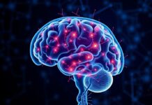
The phrase “Le rôle du cervelet dans la coordination des mouvements” invites us into a fascinating corner of neuroscience: a structure no larger than your fist that quietly conducts the orchestra of movement. You may have heard the cerebellum called the “little brain,” and while that nickname sounds cute, its responsibilities are anything but trivial. From making your cup-to-mouth motion smooth to keeping you upright while walking on a crowded street, the cerebellum is central to how we interact with the world through movement. In this article I’ll take you on a guided tour of how the cerebellum does its job, why it matters for daily life, what goes wrong when it is damaged, and what current science tells us about helping recover or enhance its function.
We will move in accessible steps: first getting to know the structure, then exploring the mechanisms of coordination, looking at clinical signs and tests, and finally examining how therapy and research are addressing cerebellar problems. I’ll keep things conversational, occasionally pausing to give concrete examples you can picture easily. Expect a mix of clear explanations, practical examples, and a couple of neat tables and lists so you can scan the essentials quickly.
Содержание
What is the cerebellum?
At a glance, the cerebellum sits under the back part of the cerebral hemispheres and behind the brainstem. It forms the rounded mass at the back of your skull just above the neck. Despite its smaller size compared to the cerebrum, it contains more neurons than the rest of the brain combined in many species — a testament to its computational intensity. The cerebellum’s surface is folded into parallel ridges, and inside it are layers of neurons arranged in circuits that are exquisitely suited to timing and error correction.
Most descriptions of the brain begin with anatomy because function follows form. The cerebellum has a distinct architecture: a cortex with specialized cells, most notably Purkinje cells, and deep cerebellar nuclei where the cortex’s output funnels to other brain areas. Purkinje cells receive an enormous number of inputs and, through their inhibitory output, shape the signals sent to the motor and premotor centers of the brain. Think of the cerebellum as an expert editor: it takes raw movement plans and sensory feedback and fine-tunes the final instructions sent to the muscles.
If you picture a control room, the cerebellum constantly receives reports about intended movement and actual movement. It compares those reports and issues corrections, often in real time, ensuring movements are precise, well-timed, and coordinated across multiple muscles and joints.
Anatomy in practical terms: zones and nuclei
Understanding the cerebellum’s subregions helps explain why different patients with cerebellar damage have different problems. Broadly, the cerebellum can be divided into three functional zones: the vestibulocerebellum, the spinocerebellum, and the cerebrocerebellum. These zones map onto anatomy and into specific deep nuclei.
The vestibulocerebellum (flocculonodular lobe) is closely tied to balance and eye movements. It communicates with vestibular nuclei to coordinate reflexes that stabilize the eyes and body during movement. The spinocerebellum (vermis and intermediate zones) handles ongoing trunk and limb coordination — it’s crucial for posture and gait. The cerebrocerebellum (lateral hemispheres) interfaces with the cerebral cortex to plan and time skilled, voluntary movements.
Within the cerebellum are the deep nuclei (fastigial, interposed, dentate). These are like relay hubs where processed information exits the cerebellum. The dentate nucleus, for example, is heavily involved in planning and initiating fine voluntary movements and communicates with motor planning areas of the cortex.
Inputs and outputs: how the cerebellum plugs into the brain
The cerebellum receives two major types of input: mossy fibers and climbing fibers. Mossy fibers bring information about sensory feedback and motor commands from many sources, while climbing fibers — originating from the inferior olive — carry powerful teaching signals that help refine cerebellar output. Within the cerebellar cortex, Purkinje cells integrate these signals and send inhibitory output to the deep nuclei. The deep nuclei then send excitatory output to motor and premotor centers, brainstem nuclei, and thalamic targets that reach the cortex.
Because of these loops — cerebellum to cortex to cerebellum — the cerebellum contributes to both feedforward control (predicting the effects of motor commands) and feedback control (correcting errors as they arise). This dual role explains why the cerebellum is essential for smooth, accurate, and adaptive movement.
How the cerebellum contributes to coordination
Coordination means that multiple muscles, joints, and sensory systems work together smoothly and efficiently to accomplish a goal. The cerebellum contributes to coordination in several interrelated ways: timing, scaling of force, sequence control, adaptation, and prediction of sensory consequences. If you’ve ever caught yourself adjusting grip when a glass slips, or corrected your step on an uneven pavement without consciously thinking about it, that’s cerebellar computation in action.
Timing is perhaps the cerebellum’s most celebrated contribution. Many movements depend on precise timing between muscle activations; for example, playing a musical instrument or typing requires microsecond-level coordination across fingers. The cerebellum helps schedule muscle contractions so that agonists and antagonists activate in the right sequence.
Scaling refers to how strongly the cerebellum influences motor output — it helps adjust the amplitude of a movement. Reach too short or overshoot a target? The cerebellum is involved in adjusting movement amplitude based on previous errors.
Sequence control means linking discrete movements into a smooth series. When you reach for a knob, twist it, and then pull — the cerebellum helps make that sequence occur fluidly.
Prediction is crucial: before sensory feedback arrives, the cerebellum predicts what the consequence of a motor command will be. These internal models let the brain issue anticipatory adjustments — for example, stiffening the shoulder when you lift a heavy suitcase, before the weight is even felt.
Feedforward versus feedback control
A useful way to think about motor control is as a balance between feedforward (predictive) and feedback (corrective) control. Feedforward control uses internal models to predict how muscles and limbs will respond and dispatches anticipatory signals. Feedback control uses sensory information — proprioception, vision, vestibular input — to correct errors after they occur. The cerebellum is bilingual: it helps build and update internal models for feedforward control and participates in fast feedback loops to correct errors rapidly.
When you reach for a coffee mug in the dark, feedforward control based on previous experience allows your hand to find the mug. If the mug is slightly moved, fast feedback mechanisms (with cerebellar involvement) let you gently adjust your grasp so you don’t spill the coffee.
Examples in everyday movement
– Gait: The rhythm and coordination between hip, knee, and ankle joints as you walk are refined by the cerebellum so steps are smooth and balanced.
– Reaching: Adjusting arm trajectory mid-reach when the target moves — the cerebellum helps correct the path with minimal delay.
– Speech: Rapid coordination of tongue, lips, and breath is partly cerebellar; damage can lead to slurred or explosive speech, known as ataxic dysarthria.
– Eye movements: The cerebellum stabilizes gaze during head movement via the vestibulo-ocular reflex and coordinates saccades (fast eye movements).
Motor learning and adaptation: the cerebellum as a teacher
One of the cerebellum’s most fascinating roles is in motor learning: the way we refine movements through practice and adapt to changes in the body or environment. Think about learning to ride a bicycle or adapting to a new pair of glasses that shift visual input. In these examples, the cerebellum helps detect errors and adjust future commands to reduce them.
At the cellular level, synaptic plasticity — especially long-term depression (LTD) of parallel fiber-Purkinje cell synapses — has been implicated in motor learning. Climbing fibers convey powerful error signals that guide changes in the strength of synapses, effectively teaching the cerebellar cortex how to respond to similar situations in the future.
Several classic experiments demonstrate cerebellar-dependent learning. In prism adaptation tasks, people wearing prism glasses that displace visual information learn to adjust their reaching movements so they reach targets accurately despite the shifted visual field. Lesions of the cerebellum impair the rate and degree of adaptation. Similarly, the adaptation of the vestibulo-ocular reflex (which stabilizes vision during head motion) requires an intact cerebellum.
From trial-and-error to smooth expertise
Early practice of a new skill often involves large, variable errors and conscious control: you think about each movement. With cerebellar learning, many of those errors shrink, sequences become smoother, and the skill becomes less attention-demanding. This transition from conscious control to automaticity is partly mediated by cerebellar-cortical circuits that shape motor programs for efficiency.
Athletes and musicians rely heavily on cerebellar learning. A pianist practicing scales repeatedly engages cerebellar mechanisms to time and coordinate finger movements precisely. As practice continues, the movements become faster, more accurate, and less consciously taxing.
Clinical signs of cerebellar dysfunction
When the cerebellum is damaged — by stroke, tumor, degenerative disease, infection, or trauma — the resulting deficits reveal its roles spectacularly. Cerebellar dysfunction typically leads to ataxia, a broad term meaning lack of coordination. Clinical signs include dysmetria (overshooting or undershooting targets), intention tremor (tremor that worsens during purposeful movement), dysdiadochokinesia (difficulty performing rapid alternating movements), gait unsteadiness, and dysarthria (slurred, scanning speech).
These signs vary depending on lesion location. Midline (vermal) lesions often cause truncal ataxia and gait problems, while lateral lesions produce limb ataxia, impaired skilled movements, and issues with motor planning. Vestibulocerebellar involvement causes dizziness, nystagmus (abnormal eye movements), and balance disturbances.
How clinicians test cerebellar function
Simple bedside tests can reveal cerebellar dysfunction:
- Finger-to-nose test: The patient touches their nose and then the examiner’s finger repeatedly; overshoot, tremor, or inaccuracy suggests dysmetria.
- Heel-to-shin test: Sliding the heel down the shin while supine; deviation or irregularity indicates lower limb cerebellar dysfunction.
- Rapid alternating movements: Rapid pronation and supination of the hands; slowed or irregular movements indicate dysdiadochokinesia.
- Gait assessment: Wide-based, unsteady gait with irregular steps suggests cerebellar ataxia; tandem gait (heel-to-toe) is particularly sensitive.
- Romberg test: While primarily a dorsal column/proprioceptive test, cerebellar patients often show difficulty in stance even with eyes open, distinguishing them from pure sensory ataxia.
These tests, together with imaging such as MRI, help localize the lesion and guide treatment.
Tables: functions, lesions, and clinical signs
Below is a concise table summarizing key cerebellar regions, their main functions, and typical clinical signs when damaged.
| Cerebellar Region | Main Functions | Typical Clinical Signs of Lesion |
|---|---|---|
| Vestibulocerebellum (flocculonodular lobe) | Balance, vestibulo-ocular reflex, eye movement coordination | Truncal instability, vertigo, nystagmus |
| Spinocerebellum (vermis & intermediate zones) | Posture, gait, limb coordination, muscle tone maintenance | Gait ataxia, limb ataxia, hypotonia |
| Cerebrocerebellum (lateral hemispheres) | Motor planning, skilled voluntary movements, motor learning | Intention tremor, dysmetria, impaired motor learning |
Another helpful table contrasts cerebellar ataxia with sensory (dorsal column) ataxia to show how clinicians differentiate them.
| Feature | Cerebellar Ataxia | Sensory (Dorsal Column) Ataxia |
|---|---|---|
| Romberg sign | Unsteady with eyes open and closed; Romberg often negative | Unsteady mainly with eyes closed; Romberg positive |
| Gait | Wide-based, irregular steps | Stomping gait, relies on visual compensation |
| Limb movements | Dysmetria, intention tremor | Loss of proprioception, difficult to judge position without vision |
Assessment and imaging
Modern neurology uses both clinical examination and imaging to evaluate cerebellar function. MRI is the gold standard to visualize cerebellar structure and identify strokes, tumors, degenerative atrophy, and inflammatory lesions. Functional imaging and electrophysiological tests can reveal altered cerebellar activity and connectivity patterns.
Genetic testing is increasingly important because many cerebellar disorders are inherited (e.g., spinocerebellar ataxias). Early identification enables counseling, prognostication, and participation in clinical trials.
When to suspect cerebellar disease
Common clinical scenarios prompting cerebellar evaluation include sudden onset of vertigo and severe imbalance (suggesting stroke), progressive worsening of coordination over months to years (suggesting degenerative disease), or subacute cerebellar symptoms after infection or exposure to toxins or medications. Alcohol and certain chemotherapeutic agents can damage cerebellar neurons, causing ataxia.
Rehabilitation and treatment strategies
While some cerebellar damage is irreversible, many patients improve with targeted therapies. Rehabilitation focuses on retraining coordination, improving balance, and teaching compensatory strategies. Physical and occupational therapists design exercises that challenge balance, promote step and trunk control, and refine limb coordination. Repetition and task-specific practice leverage residual cerebellar plasticity and alternative neural pathways to regain function.
Evidence supports benefits from:
- Task-oriented training: Practicing specific tasks (walking, reaching) improves performance more than nonspecific exercise.
- Balance training: Exercises that progressively challenge posture and vestibular control.
- Assistive devices: Canes, orthoses, or adaptive utensils that reduce fall risk and support independence.
- Speech therapy: For dysarthria, therapy helps with pacing, breath control, and articulation strategies.
Emerging techniques that show promise include noninvasive brain stimulation (transcranial magnetic stimulation, transcranial direct current stimulation) aimed at modulating cerebellar-cortical circuits, and pharmacological agents that target neurotransmitter systems involved in cerebellar circuitry. Results are preliminary but intriguing: combining stimulation with task practice may enhance motor learning in some patients.
Neurosurgical and disease-specific approaches
When cerebellar dysfunction is caused by a structurally correctable lesion such as a tumor, surgical removal can restore or preserve functions. For degenerative conditions, symptomatic treatments and multidisciplinary management remain the mainstay. In rare cases of immune-mediated cerebellar ataxia, immunotherapy can be effective if started early.
Rehabilitation outcomes vary. Younger patients and those with focal lesions tend to recover more function than those with diffuse degenerative disease. Still, even in progressive disorders, therapy often improves safety and quality of life.
Beyond movement: cognitive and emotional roles of the cerebellum
For many years the cerebellum was thought to be purely motor. Over the past decades, research has shown it also contributes to cognition, language, and emotion. Patients with cerebellar lesions can develop the “cerebellar cognitive affective syndrome,” which includes impairments in executive function, spatial processing, language, and affective regulation. This expanded view positions the cerebellum as a general-purpose predictive and timing engine that the brain employs not just for muscles, but for thought and feeling as well.
Functional imaging shows cerebellar activation during working memory tasks, language, and even social cognition. The same principles of timing and prediction that help coordinate muscles may help synchronize mental operations across different brain areas.
Why this matters clinically
Acknowledging cognitive and emotional roles changes how clinicians approach cerebellar patients. Evaluation often includes cognitive screening and mood assessment. Rehabilitation may incorporate cognitive exercises and strategies to manage executive dysfunction. Recognizing these non-motor symptoms improves holistic care and patient outcomes.
Development, aging, and plasticity
The cerebellum develops early in life and is critical for motor milestones such as sitting, crawling, and walking. In childhood, cerebellar damage can cause long-lasting motor and cognitive effects, but the young brain also has remarkable plasticity. Early intervention and therapy can yield substantial gains.
With aging, cerebellar volume and function decline, contributing to slower movements and increased fall risk. Distinguishing normal aging from pathological cerebellar degeneration is important for management. Lifestyle factors like physical activity appear to help maintain cerebellar function by promoting motor learning and balance.
Research frontiers: what scientists are learning now
Current research explores how cerebellar circuits implement prediction and learning at cellular resolution, how the cerebellum interacts with other brain networks, and how noninvasive stimulation can enhance rehabilitation. Genetic studies are unraveling the many subtypes of spinocerebellar ataxia, opening paths to targeted therapies. Other exciting areas include brain–computer interfaces that might bypass damaged cerebellar pathways and artificial intelligence models inspired by cerebellar computation.
Animal studies continue to reveal principles of cerebellar computation, and human trials are testing novel rehabilitation protocols that integrate virtual reality, robotics, and neuromodulation. Researchers are also investigating how cerebellar dysfunction contributes to neuropsychiatric conditions like autism and schizophrenia, potentially offering new therapeutic targets.
Practical takeaways for patients, families, and caregivers
If you or a loved one has cerebellar dysfunction, here are practical points to keep in mind:
- Early assessment matters. Timely diagnosis can identify reversible causes and allow early rehabilitation.
- Regular, task-specific practice helps. Repetition of meaningful tasks encourages motor learning and functional gains.
- Fall prevention is essential. Home modifications, assistive devices, and balance training reduce risk.
- Address speech and swallowing issues early. Speech therapy improves communication and reduces aspiration risk.
- Support cognition and mood. Cognitive and emotional changes are real and deserve attention alongside motor symptoms.
These are practical, actionable strategies that complement medical and surgical care.
Common misconceptions
It helps to clear up a few common misunderstandings about the cerebellum:
- Misconception: The cerebellum only controls balance. Reality: It handles timing, coordination, motor learning, and contributes to cognition and emotion.
- Misconception: Damage to the cerebellum always causes weakness. Reality: Cerebellar problems usually cause incoordination, not primary muscle weakness.
- Misconception: Recovery from cerebellar injury is impossible. Reality: Rehabilitation and plasticity often produce meaningful improvements, especially with early and focused therapy.
Understanding these nuances can help patients and families set realistic expectations and focus on therapies that improve daily function.
Summary of mechanisms in a quick list
For quick reference, here are the cerebellum’s core mechanisms that support movement:
- Integration of sensory inputs and motor plans to align intended and actual movement.
- Timing and sequencing of muscle activation patterns for smooth motion.
- Error detection via climbing fiber signals and synaptic plasticity to drive motor learning.
- Adjustment of motor output amplitude and coordination across joints and limbs.
- Feedforward prediction of sensory consequences to enable anticipatory adjustments.
These mechanisms interact continuously to produce the graceful, adaptive movements we often take for granted.
Conclusion
The cerebellum, central to “Le rôle du cervelet dans la coordination des mouvements,” is a compact but powerful structure whose contributions to human movement are essential and multifaceted: it times, scales, predicts, and learns, constantly refining the commands that let us reach, walk, speak, and react with fluid precision; when it malfunctions we see dramatic but sometimes treatable disruptions in balance, coordination, and even thought, and ongoing research and rehabilitation strategies offer hope for restoring function and improving quality of life.

